Submitted by Laura Puhl on Thu, 21/11/2024 - 11:32
This week marks a milestone for the Human Cell Atlas project, a consortium of researchers co-led by CSCI's Professor Sarah Teichmann and Dr Aviv Regev of Genentech.
A collection of 13 papers in Nature have been published this week revealing data from embryonic, fetal and paediatric organs, as well as computational tools, which provide a foundation for the first draft of a full Human Cell Atlas. Alongside publications released earlier in the year and a few more ahead, these papers are released as a full collection of 43 new papers in the ongoing mission to complete a map of each of the 37 trillion cells in the human body.
Human Cell Atlas
The Human Cell Atlas (HCA) was founded in 2016 at the Wellcome Trust in London, and now numbers more than 3,600 members in over 100 countries with the shared goal of a completed map of human cells. Researchers aim to have a first draft of the atlas done in the next year or two, hoping that intricate knowledge of human cells can be used for diagnostic and therapeutic purposes.
Using new and developing genomics technologies, researchers are working to map the 'molecular fingerprint' of each cell in order to eventually create a readable map of the body. Researchers involved in these studies are now able, with new technologies, to measure cells at single cell resolution, allowing them to look closer at different cell types and more closely monitor what they do. This has important implications for diagnostics and drug discovery, as well as contributing to a more detailed understanding of the human body.
Professor Teichmann says, 'Having a healthy human map for large cohorts of people helps us understand what the cells are like in their normal ground state or reference state and then provide that framework for comparison to disease samples. It allows us to study the mechanisms of disease and to understand progressions of disease better for drug discovery.'
Professor Muzlifah Haniffa, Affiliate PI of CSCI and member on the HCA organising committee, says, "Fundamentally, these studies tell us how tissues, organs and humans are built. Understanding human development is critical to understand developmental disorders, childhood disorders that have a prenatal onset, as well as diseases that also affect adults, as developmental pathways can re-emerge in later life disease. Practical applications include new diagnostic, clinical management and therapeutic strategies for the clinic."
New Research Highlights
|
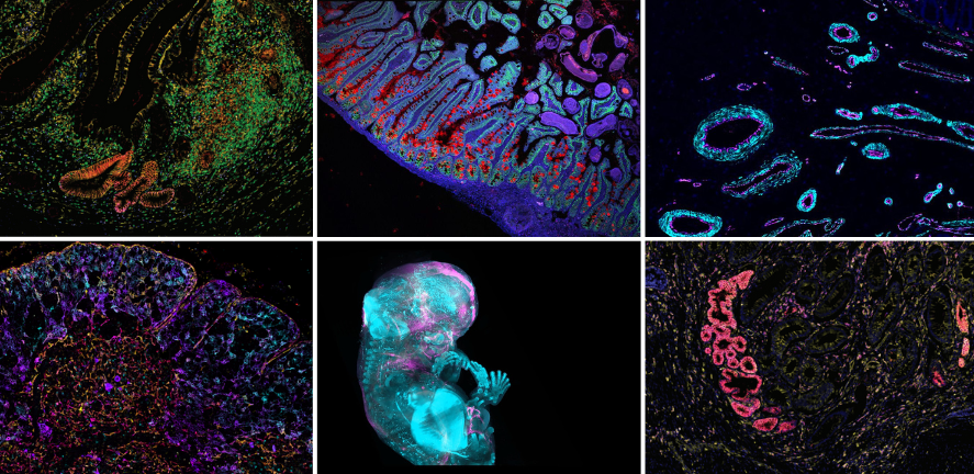
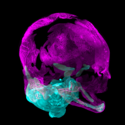
Early skeleton map reveals how bones form in humans during the first trimester The study shows a clear picture of how cartilage acts as a scaffold for bone development across the skeleton, apart from the top of the skull. This sheds light on the process of arthritis and highlights cells involved in skull and bone growth. Image credit: A.Chédotal & R. Blain, Institut de la Vision, Paris & MeLiS/UCBL/ HCL, Lyon |
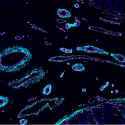
Detailed map of human blood vessel cells could transform treatment of diseases In a significant advance, scientists have for the first time identified and mapped the different types of blood vessels in the human body, finding 42 distinct types of vascular cells in 19 organs and tissues. All of the data from the Human Vascular Cell Atlas study has been made available on an open-access, interactive portal, so that the world’s scientists and researchers can benefit. Image credit: Ana-Maria Cujba (Teichmann Lab)
|
|
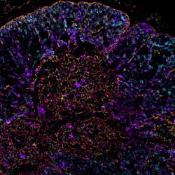
1.6 million gut cells mapped to find new ways to treat bowel diseases Researchers combined datasets to create the most comprehensive cell map of the human gut, offering insights into bowel conditions and possible treatments. Image credit: A Oliver |
Map of human thymus shows development of immune responses in early life Researchers have uncovered key differences in the development of immune cells, offering insight to therapies for those with compromised immune systems. Image credit: Andrea Radtke NIH |


